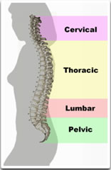
Back pain (also known "dorsalgia") is pain felt in the back that usually originates from the muscles, nerves, bones, joints or other structures in the spine.
The pain may have a sudden onset or can be a chronic pain; it can be constant or intermittent, stay in one place or radiate to other areas. It may be a dull ache, or a sharp or piercing or burning sensation. The pain may be felt in the neck (and might radiate into the arm and hand), in the upper back, or in the low back, (and might radiate into the leg or foot), and may include symptoms other than pain, such as weakness, numbness or tingling.
Back pain is one of humanity's most frequent complaints. In the U.S., acute low back pain (also called lumbago) is the fifth most common reason for physician visits. About nine out of ten adults experience back pain at some point in their life, and five out of ten working adults have back pain every year.
The spine is a complex interconnecting network of nerves, joints, muscles, tendons and ligaments, and all are capable of producing pain. Large nerves that originate in the spine and go to the legs and arms can make pain radiate to the extremities.
Associated conditions
Back pain can be a sign of a serious medical problem, although this is not most frequently the underlying cause
»
Typical warning signs of a potentially life-threatening problem are bowel and/or bladder incontinence or progressive weakness in the legs.
»
Severe back pain (such as pain that is bad enough to interrupt sleep) that occurs with other signs of severe illness (e.g. fever, unexplained weight loss) may also indicate a serious underlying medical condition.
»
Back pain that occurs after a trauma, such as a car accident or fall may indicate a bone fracture or other injury.
»
Back pain in individuals with medical conditions that put them at high risk for a spinal fracture, such as osteoporosis or multiple myeloma, also warrants prompt medical attention.
Back pain does not usually require immediate medical intervention. The vast majority of episodes of back pain are self-limiting and non-progressive. Most back pain syndromes are due to inflammation, especially in the acute phase, which typically lasts for two weeks to three months.
A few observational studies suggest that two conditions to which back pain is often attributed, lumbar disc herniation and degenerative disc disease may not be more prevalent among those in pain than among the general population and that the mechanisms by which these conditions might cause pain are not known. Other studies suggest that for as many as 85% of cases, no physiological cause can be shown.
A few studies suggest that psychosocial factors such as on-the-job stress and dysfunctional family relationships may correlate more closely with back pain than structural abnormalities revealed in x-rays and other medical imaging scans.
Underlying causes
Muscle strains (pulled muscles) are commonly identified as the cause of back pain, as are muscle imbalances. Pain from such an injury often remains as long as the muscle imbalances persist. The muscle imbalances cause a mechanical problem with the skeleton, building up pressure at points along the spine, which causes the pain.
Another cause of acute low back pain is a meniscoid occlusion. The more mobile regions of the spine, such as the facet joints, have invaginations of their synovial membranes that act as a cushion to help the bones move over each other smoothly. The synovial membrane is well supplied with blood and nerves. When these become pinched or trapped sudden severe pain may result. The pinching causes the membrane to become inflamed, causing greater pressure and ongoing pain. Symptoms include severe low back pain that may be accompanied by muscle spasm, pain with walking, concentration of pain to one side, but no radiculopathy (radiating pain down buttock and leg). Relief should be felt with flexion (bending forward),and exacerbated with extension (bending backward).
When back pain lasts more than three months, or if there is more radicular pain (sciatica) than back pain, a more specific diagnosis can usually be made. There are several common causes of back pain: for adults under age 50, these include spinal disc herniation and degenerative disc disease or isthmic spondylolisthesis; in adults over age 50, common causes also include osteoarthritis (degenerative joint disease) and spinal stenosis,trauma, cancer, infection, fractures, and inflammatory disease[1]. Non-anatomical factors can also contribute to or cause back pain, such as stress,[12] repressed anger,[13] or depression. Even if there is an anatomical cause for the pain, if depression is present it should also be treated concurrently.
New attention has been focused on non-discogenic back pain, where patients have normal or near-normal MRI and CT scans. One of the newer investigations looks into the role of the dorsal ramus in patients that have no radiographic abnormalities. See Posterior Rami Syndrome.
Treatment
The management goals when treating back pain are to achieve maximal reduction in pain intensity as rapidly as possible; to restore the individual's ability to function in everyday activities; to help the patient cope with residual pain; to assess for side-effects of therapy; and to facilitate the patient's passage through the legal and socioeconomic impediments to recovery. For many, the goal is to keep the pain to a manageable level to progress with rehabilitation, which then can lead to long term pain relief. Also, for some people the goal is to use non-surgical therapies to manage the pain and avoid major surgery, while for others surgery may be the quickest way to feel better.
Not all treatments work for all conditions or for all individuals with the same condition, and many find that they need to try several treatment options to determine what works best for them. The present stage of the condition (acute or chronic) is also a determining factor in the choice of treatment. Only a minority of back pain patients (most estimates are 1% - 10%) require surgery.
Neck pain
Neck pain (or cervicalgia) is a common problem, with two-thirds of the population having neck pain at some point in their lives.[1] It is increasing in both intensity, frequency and severity of episodes.[citation needed] As people are increasingly sedentary, live fast-paced and hectic lives, they place more stress and strain on the upper back and neck regions of their spines.[citation needed]
Neck pain, although felt in the neck, can be caused by numerous other spinal issues. Neck pain may arise due to muscular tightness in both the neck and upper back. Joint disruption in the neck creates pain, as does joint disruption in the upper back.
The head is supported by the lower neck and upper back, and it is these areas that commonly cause neck pain. The top three joints in the neck allow for most movement of your neck and head. The lower joints in the neck and those of the upper back create a supportive structure for your head to sit on. If this support system is affected adversly, then the muscles in the area will tighten, leading to neck pain.
Neck pain may also arise from many other physical and emotional health issues.
Neck Pain Causes
Reasons for neck pain can be complex. Major and severe causes of neck pain include:
»
Spondylosis - degenerative arthritis and osteophytes
»
Spinal stenosis – a narrowing of the spinal canal
»
Spinal disc herniation – protruding or bulging discs, or if severe prolapse.
»
Severe degeneration – usually as a result of past injuries or whiplash accidents.
The more common and lesser neck pain causes include
»
Stress – physical and emotional stresses
»
Prolonged postures – many people fall asleep on sofas and chairs and wake with sore necks
»
Minor injuries and falls – car accidents, sporting events and day to day minor injuries
»
Referred pain – mostly from upper back problems
»
Over-use – muscular strain is one of the most common causes
Although the causes are numerous, most are easily recitified by either professional help or using self help advice and techniques.
Treatment
Neck pain is treated by numerous physical therapies. They range in complexity depending on the severity and underlying causes of the pain. Treatment is administered by chiropractic, osteopathic and physical therapy. All of these specialties treat neck pain issues. The benefit of mobilisation and manipulation is not clear. Neck pain can also be eased via many self help techniques such as stretching, strength building exercises. Non-traditional methods such as Acupressure, Reflexology
Ostho Arthritis
Arthritis (from Greek arthro-, joint + -itis, inflammation; plural: arthritides) is a group of conditions involving damage to the joints of the body. Arthritis is the leading cause of disability in people older than fifty-five years.
There are different forms of arthritis; each has a different cause. The most common form of arthritis, osteoarthritis (degenerative joint disease) is a result of trauma to the joint, infection of the joint, or age. Emerging evidence suggests that abnormal anatomy might contribute to the early development of osteoarthritis. Other arthritis forms are rheumatoid arthritis and psoriatic arthritis, autoimmune diseases in which the body attacks itself. Septic arthritis is caused by joint infection. Gouty arthritis is caused by deposition of uric acid crystals in the joint, causing inflammation. There is also an uncommon form of gout caused by the formation of rhomboid crystals of calcium pyrophosphate. This gout is known as pseudogout.
Blood tests and X-rays of the affected joints often are performed to make the diagnosis. Screening blood tests are indicated if certain arthritides are suspected. These might include: rheumatoid factor, antinuclear factor (ANF), extractable nuclear antigen, and specific antibodies.
Types of arthritis
Primary forms of arthritis
»
Osteoarthritis
»
Rheumatoid arthritis
»
Septic arthritis
»
Gout and pseudogout
»
Juvenile idiopathic arthritis
»
Still's disease
»
Ankylosing spondylitis
Secondary to other diseases
»
Lupus erythematosus
»
Henoch-Schönlein purpura
»
Psoriatic arthritis
»
Reactive arthritis
»
Haemochromatosis
»
Hepatitis
»
Wegener's granulomatosis (and many other vasculitis syndromes)
»
Lyme disease
»
Familial Mediterranean fever
»
Hyperimmunoglobulinemia D with recurrent fever
»
TNF receptor associated periodic syndrome
»
Inflammatory bowel disease (Including Crohn's Disease and Ulcerative Colitis)
Diseases that can mimic arthritis include
»
Hypertrophic osteoarthropathy
»
Multiple myeloma
»
Osteoporosis
»
Fifth disease
Treatment
Treatment options vary depending on the type of arthritis and include physical and occupational therapy, lifestyle changes (including exercise and weight control), medications and dietary supplements (symptomatic or targeted at the disease process causing the arthritis). Arthroplasty (joint replacement surgery) may be required in eroding forms of arthritis.
In general, studies have shown that physical exercising of the affected joint can have noticeable improvement in terms of long-term pain relief. Furthermore, exercising of the arthritic joint is encouraged to maintain the health of the particular joint and the overall body of the person.
Another form of non-drug treatment that does have a body of proper research to support its efficacy is marine oil, from both fish and the New Zealand green-lipped mussel (Perna canaliculus). Diets high in marine oils from cold-water fish such as salmon, mackerel, and tuna have been shown to reduce the inflammation of joint conditions such as arthritis.
History
While evidence of primary ankle (kaki) osteoarthritis has been discovered in dinosaurs, the first known traces of human arthritis date back as far as 4500 BC. It was noted in skeletal remains of Native Americans found in Tennessee and parts of what is now Olathe, Kansas. Evidence of arthritis has been found throughout history, from Ötzi, a mummy (circa 3000 BC) found along the border of modern Italy and Austria, to the Egyptian mummies circa 2590 BC.
Rheumatoid arthritis
Rheumatoid arthritis (RA) is a chronic, systemic autoimmune disorder that causes the immune system to attack the joints, causing inflammation (arthritis), and some organs, such as the lungs and skin. It can be a disabling and painful condition, which can lead to substantial loss of functioning mobility due to pain and joint destruction. It is diagnosed with blood tests (especially a test called rheumatoid factor) and X-rays. Diagnosis and long-term management are typically performed by a rheumatologist, an expert in the diseases of joints and connective tissues.
Various treatments are available. Non-pharmacological treatment includes physical therapy and occupational therapy. Analgesia (painkillers) and anti-inflammatory drugs, as well as steroids, are used to suppress the symptoms, while disease-modifying antirheumatic drugs (DMARDs) are often required to reverse the disease process and prevent long-term damage. Classic DMARDs are methotrexate and sulfasalazine, but also the newer group of biologics which includes highly-effective agents such as infliximab (Remicade), etanercept (Embrel), adalimumab (Humira), abatacept (Orencia) and rituximab (Rituxan/Mabthera).
The name is based on the term "rheumatic fever", an illness which includes joint pain and is derived from the Greek word rheumatos ("flowing"). The suffix -oid ("resembling") gives the translation as joint inflammation that resembles rheumatic fever. The first recognized description of rheumatoid arthritis was made in 1800 by Dr Augustin Jacob Landré-Beauvais (1772-1840) of Paris.
Signs and symptoms
While rheumatoid arthritis primarily affects joints, problems involving all other organs of the body are known to occur. Extra-articular ("outside the joints") manifestations occur in about 15% of individuals with rheumatoid arthritis.[3] It can be difficult to determine whether disease manifestations are directly caused by the rheumatoid process itself, or from side effects of the medications commonly used to treat it - for example, lung fibrosis from methotrexate, or osteoporosis from corticosteroids.
Joints
The arthritis of rheumatoid arthritis is due to synovitis, which is inflammation of the synovial membrane that covers the joint. Joints become red, swollen, tender and warm, and stiffness prevents their use. By definition, RA affects multiple joints (it is a polyarthritis). Most commonly, small joints of the hands, feet and cervical spine are affected, but larger joints like the shoulder and knee can also be involved, differing per individual. Eventually, synovitis leads to erosion of the joint surface, causing deformity and loss of function.
Inflammation in the joints manifests itself as a soft, "doughy" swelling, causing pain and tenderness to palpation and movement, a sensation of localised warmth, and restricted movement. Increased stiffness upon waking is often a prominent feature and may last for more than an hour. These signs help distinguish rheumatoid from non-inflammatory diseases of the joints such as osteoarthritis (sometimes referred to as the "wear-and-tear" of the joints). In RA, the joints are usually affected in a fairly symmetrical fashion although the initial presentation may be asymmetrical.
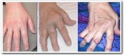
As the pathology progresses the inflammatory activity leads to erosion and destruction of the joint surface, which impairs their range of movement and leads to deformity. The fingers are typically deviated towards the little finger (ulnar deviation) and can assume unnatural shapes. Common deformities in rheumatoid arthritis are the Boutonniere deformity (Hyperflexion at the proximal interphalangeal joint with hyperextension at the distal interphalangeal joint), swan neck deformity (Hyperextension at the proximal interphalangeal joint, hyperflexion at the distal interphalangeal joint). The thumb may develop a "Z-Thumb" deformity with fixed flexion and subluxation at the metacarpophalangeal joint, and hyperextension at the IP joint.
Hands affected by RA
Fracture
A fracture is the (local) separation of an object or material into two, or more, pieces under the action of stress.
The word fracture is often applied to bones of living creatures, or to crystals or crystalline materials, such as gemstones or metal. Sometimes, in crystalline materials, individual crystals fracture without the body actually separating into two or more pieces. Depending on the substance which is fractured, a fracture reduces strength (most substances) or inhibits transmission of light (optical crystals).
A detailed understanding of how fracture occurs in materials may be assisted by the study of fracture mechanics.
Types of fracture
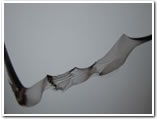
Brittle fracture
Brittle fracture in glass.
Fracture of an Aluminum Crank Arm. Bright: Brittle fracture. Dark: Fatigue fracture.
In brittle fracture, no apparent plastic deformation takes place before fracture. In brittle crystalline materials, fracture can occur by cleavage as the result of tensile stress acting normal to crystallographic planes with low bonding (cleavage planes). In amorphous solids, by contrast, the lack of a crystalline structure results in a conchoidal fracture, with cracks proceeding normal to the applied tension.
The theoretical strength of a crystalline material is (roughly)
where
E is the Young's modulus of the material,
γ is the surface energy, and
ro is the equilibrium distance between atomic centers.
On the other hand, a crack introduces a stress concentration modeled by
(For sharp cracks)
where
σapplied is the loading stress,
a is half the length of the crack, and
ρ is the radius of curvature at the crack tip.
Putting these two equations together, we get
Looking closely, we can see that sharp cracks (small ρ) and large defects (large a) both lower the fracture strength of the material.
Recently, scientists have discovered supersonic fracture , the phenomenon of crack motion faster than the speed of sound in a material.[citation needed] This phenomenon was recently also verified by experiment of fracture in rubber-like materials.
Ductile fracture
Ductile failure of a specimen strained axially.
Schematic representation of the steps in ductile fracture.
In ductile fracture, extensive plastic deformation takes place before fracture. Many ductile metals, especially materials with high purity, can sustain very large deformation of 50–100% or more strain before fracture under favorable loading condition and environmental condition. The strain at which the fracture happens is controlled by the purity of the materials. At room temperature, pure iron can undergo deformation up to 100% strain before breaking, while cast iron or high-carbon steels can barely sustain 3% of strain.[citation needed].
Because ductile rupture involves a high degree of plastic deformation, the fracture behavior of a propagating crack as modeled above changes fundamentally. Some of the energy from stress concentrations at the crack tips is dissipated by plastic deformation before the crack actually propagates.
The basic steps of ductile fracture are necking (which results in stress localization at the point on the sample of smallest cross-sectional area), void formation, void coalescence (also known as crack formation), crack propagation, and failure, often resulting in a cup-and-cone shaped failure surface.
Crack Separation Modes
The three fracture modes.
There are three ways of applying a force to enable a crack to propagate
»
Mode I crack – Opening mode (a tensile stress normal to the plane of the crack)
»
Mode II crack – Sliding mode (a shear stress acting parallel to the plane of the crack and perpendicular to the crack front)
»
Mode III crack – Tearing mode (a shear stress acting parallel to the plane of the crack and parallel to the crack front)
Dislocation
Occurrence
Although it is possible for any joint to become subluxed or dislocated, the most common sites it is seen in the human body are
»
Shoulders,
»
Fingers,
»
Knees,
»
Wrists (most likely be accompanied by a fracture.)
»
Elbows (most likely be accompanied by a fracture.)
Treatment
Anyone experiencing a joint dislocation or subluxation should seek medical assistance as soon as possible. A dislocated joint can only be successfully 'reduced' into its normal position by a trained medical professional. Trying to reduce a joint without any training could result in making the injury substantially worse.
X-rays are usually taken to confirm a diagnosis and detect any fractures which may also have occurred at the time of dislocation.
Once a diagnosis is confirmed, the joint is usually manipulated back into position. This can be a very painful process, therefore this is typically done either in A&E under sedation or in an Operating Room under a General anaesthetic.
It is important the joint is reduced as soon as possible, as in the state of dislocation, the blood supply to the joint (or distal anatomy) may be compromised. This is especially true in the case of a dislocated ankle, due to the anatomy of the blood supply to the foot.
Shoulder injuries can also be surgically stabilized, depending on the severity, using arthroscopic surgery.
Some joints are more at risk of becoming dislocated again after an initial injury. This is due to the weakening of the muscles and ligaments which hold the joint in place. Shoulders a prime example of this. Any shoulder dislocation should be followed up with thorough physiotherapy.
There are some medical conditions by where joint dislocations are frequent and spontaneous, such as Ehlers-Danlos Syndrome and Congenital Hip Dysplasia.
Osteoporosis
Osteoporosis is a disease of bone that leads to an increased risk of fracture. In osteoporosis the bone mineral density (BMD) is reduced, bone microarchitecture is disrupted, and the amount and variety of non-collagenous proteins in bone is altered. Osteoporosis is defined by the World Health Organization (WHO) in women as a bone mineral density 2.5 standard deviations below peak bone mass (20-year-old healthy female average) as measured by DXA; the term "established osteoporosis" includes the presence of a fragility fracture.[1] Osteoporosis is most common in women after menopause, when it is called postmenopausal osteoporosis, but may also develop in men, and may occur in anyone in the presence of particular hormonal disorders and other chronic diseases or as a result of medications, specifically glucocorticoids, when the disease is called steroid- or glucocorticoid-induced osteoporosis (SIOP or GIOP). Given its influence on the risk of fragility fracture, osteoporosis may significantly affect life expectancy and quality of life.
Osteoporosis can be prevented with lifestyle advice and sometimes medication, and in people with osteoporosis treatment may involve lifestyle advice, preventing falls and medication (calcium, vitamin D, bisphosphonates and several others).
Signs and symptoms
Osteoporosis itself has no specific symptoms; its main consequence is the increased risk of bone fractures. Osteoporotic fractures are those that occur in situations where healthy people would not normally break a bone; they are therefore regarded as fragility fractures. Typical fragility fractures occur in the vertebral column, rib, hip and wrist.
Fractures
The symptoms of a vertebral collapse ("compression fracture") are sudden back pain, often with radiculopathic pain (shooting pain due to nerve compression ) and rarely with spinal cord compression or cauda equina syndrome. Multiple vertebral fractures lead to a stooped posture, loss of height, and chronic pain with resultant reduction in mobility.
Fractures of the long bones acutely impair mobility and may require surgery. Hip fracture, in particular, usually requires prompt surgery, as there are serious risks associated with a hip fracture, such as deep vein thrombosis and a pulmonary embolism, and increased mortality.
Falls risk
The increased risk of falling associated with aging leads to fractures of the wrist, spine and hip. The risk of falling, in turn, is increased by impaired eyesight due to any cause (e.g. glaucoma, macular degeneration), balance disorder, movement disorders (e.g. Parkinson's disease), dementia, and sarcopenia (age-related loss of skeletal muscle). Collapse (transient loss of postural tone with or without loss of consciousness) leads to a significant risk of falls; causes of syncope are manifold but may include cardiac arrhythmias (irregular heart beat), vasovagal syncope, orthostatic hypotension (abnormal drop in blood pressure on standing up) and seizures. Removal of obstacles and loose carpets in the living environment may substantially reduce falls. Those with previous falls, as well as those with a gait or balance disorder, are most at risk.
Joint Replacement
Joint replacement is one of the most common and successful operations in modern orthopaedic surgery. It consists of replacing painful, arthritic, worn or cancerous parts of the joint with artificial surfaces shaped in such a way as to allow joint movement.
Prognosis is good to excellent in 95% of major joint replacements (hips and knees). Pain relief is especially reliable. Full recovery of range of motion is not always accomplished.
Pre-operative work-up
Because of the major surgery a complete pre-anaesthetic work-up is required. In elderly patients this usually would include ECG, Chest Xray, urine tests, haematology and biochemistry blood tests. Cross match of blood is routine also as a high percentage of patients receive a blood transfusion. Pre-operative planning requires accurate Xrays of the affected joint. The implant design is selected and the size matched to the xray images (a process known as templating).
Recovery
A few days hospitalization followed by several weeks of protected function, healing and rehabilitation. This may then be followed by several months of slow improvement in strength and endurance.
Post-operative rehabilitation
Early mobilisation of the patient is thought to be the key to reducing the chances of complications such as venous thromboembolism and Pneumonia. Modern practice is to mobilize patients as soon as possible and ambulate with walking aids when tolerated. Depending on the joint involved and the pre-op status of the patient the time of hospitalization varies from 1 day to 2 weeks with the average being 4-7 days in most regions.
Physiotherapy is used extensively to help patients recover function after joint replacement surgery. A graded exercise programme is needed. Initially the patients' muscles have not healed after the surgery; exercises for range of motion of the joints and ambulation should not be strenuous. Later when the muscle is healed the aim of exercise expands to include strengthening and recovery of function.
Arthroscopy
Arthroscopy (also called arthroscopic surgery) is a minimally invasive surgical procedure in which an examination and sometimes treatment of damage of the interior of a joint is performed using an arthroscope, a type of endoscope that is inserted into the joint through a small incision. Arthroscopic procedures can be performed either to evaluate or to treat many orthopaedic conditions including torn floating cartilage, torn surface cartilage, ACL reconstruction, and trimming damaged cartilage.
The advantage of arthroscopy over traditional open surgery is that the joint does not have to be opened up fully. Instead, only two small incisions are made - one for the arthroscope and one for the surgical instruments. This reduces recovery time and may increase the rate of surgical success due to less trauma to the connective tissue. It is especially useful for professional athletes, who frequently injure knee joints and require fast healing time. There is also less scarring, because of the smaller incisions. Irrigation fluid is used to distend the joint and make a surgical space. Sometimes this fluid leaks into the surrounding soft tissue causing extravasation and edema
The surgical instruments used are smaller than traditional instruments. Surgeons view the joint area on a video monitor, and can diagnose and repair torn joint tissue, such as ligaments and menisci.
Arthroscopy is used for joints of the knee, shoulder, elbow, wrist, ankle, and hip.
Knee arthroscopy
Lateral meniscus located between thigh bone (femur, above) and shin bone (tibia, below). The tibial cartilage displays a fissure (tip of teaser instrument).
Knee arthroscopy has in many cases replaced the classic arthrotomy that was performed in the past. Today knee arthroscopy is commonly performed for treating meniscus injury, reconstruction of the anterior cruciate ligament and for cartilage microfracturing. Arthroscopy can also be performed just for diagnosing and checking of the knee; however, the latter use has been mainly replaced by magnetic resonance imaging.
During an average knee arthroscopy, a small fiberoptic camera (the endoscope) is inserted into the joint through a small incision, about 4 mm (1/8 inch) long. A special fluid is used to visualize the joint parts. More incisions might be performed in order to check other parts of the knee. Then other miniature instruments are used and the surgery is performed.
Recovery after a knee arthroscopy is a lot faster compared to arthrotomy. Most patients can return home and walk using crutches the same or the next day after the surgery. Recovery time depends on the reason that surgery was needed and the patient's physical condition. Usually a patient can fully load his leg already within a couple of days and after a few weeks the joint function can fully recover. It is not uncommon for athletes who have a beyond average physical condition to return to normal athletic activities within a few weeks.
Arthroscopic surgeries of the knee are done for many reasons, but the usefulness of surgery for treating osteoarthritis is doubtful. A double-blind placebo-controlled study on arthroscopic surgery for osteoarthritis of the knee was published in the New England Journal of Medicine in 2002.[2] In this three-group study, 180 military veterans with osteoarthritis of the knee were randomly assigned to receive arthroscopic débridement with lavage, just arthroscopic lavage, or a sham surgery, which made superficial incisions to the skin while pretending to do the surgery. For two years after the surgeries, patients reported their pain levels and were evaluated for joint motion. Neither the patients nor the independent evaluators knew which patients had received which surgery. The study reported, "At no point did either of the intervention groups report less pain or better function than the placebo group."[3] Because there is no confirmed usefulness for these surgeries, many agencies are reconsidering paying for a surgery which seems to create risks with no benefit.
Spinal arthroscopy
Many invasive spine procedures involve the removal of bone, muscle, and ligaments to access and treat problematic areas. In some cases, thoracic (mid-spine) conditions requires a surgeon to access the problem area through the rib cage, dramatically lengthening recovery time.
Arthroscopic (also endoscopic) spinal procedures allow a surgeon to access and treat a variety of spinal conditions with minimal damage to surrounding tissues. Recovery times are greatly reduced due to the relatively small size of incision(s) required, and many patients are treated on an outpatient basis.[5] Recovery rates and times vary according to condition severity and the patient's overall health.
Arthroscopic procedures treat
»
Spinal disc herniation and degenerative discs
»
spinal deformity
»
tumors
»
general spine trauma
Wrist arthroscopy
Arthroscopic view showing two of the wrist bones.
Arthroscopy of the wrist is used to investigate and treat symptoms of repetitive strain injury, fractures of the wrist and torn or damaged ligaments. It can also be used to ascertain joint damage caused by arthritis.
Minimally Invasive Spine Surgery
PATIENTS PREFER MINIMALLY invasive techniques because such techniques reduce recovery times and provide cosmetic benefits. Reviewing the history of minimally invasive surgery helps us understand the advances in spine surgery. Minimally invasive spine surgery has adopted techniques from several fields to better treat spinal disorders. Minimally invasive spine surgery has been influenced by advances in lasers, endoscopy, and image guidance systems. Discogenic disorders have been treated by using chemonucleolysis, automated percutaneous discectomy, and intradiscal thermoablation. Endoscopic techniques have been used to treat spinal disorders. Thoracoscopes and laparoscopes have been used to perform anterior release of scoliotic or kyphotic deformities and to perform transthoracic microsurgical discectomies. The role of spinal thoracoscopy has expanded to include corpectomy, vertebral reconstruction with internal fixation, hardware application, and resection of neurogenic, spinal, and paraspinal tumors. Advances in interbody fusion cage technology have generated a great deal of interest in laparoscopic techniques. Image-guided systems are widely used in intracranial surgery and have been adapted to facilitate screw placement since the middle 1990s. The use of image-guided systems for pedicle screw placement has improved placement accuracy. The system relies on precise localization of the pedicles with computed tomography. Minimally invasive surgery is designed for "conventional" operations involving extensive anatomic dissections performed via small incisions; it yields shorter recovery times and less morbidity.
Foot
The human foot is of the plantigrade form. The bottom of the foot is called the sole and the area just behind the toes is called the ball. The skin at the sole of the foot is denser than any other skin on the human body. The evolution of man has seen the density of the sole of the foot increase as man developed the ability to walk using the legs only.
Bones
The bones of the human foot
The major bones in the human foot are
»
Phalanges: The bones in the toes are called phalanges.
»
Metatarsals: The bones in the middle of the foot are called metatarsal bones.
»
Cuneiforms: There are three bones in the middle of the foot, towards the centre of the body, called cuneiforms.
»
Cuboid: The bone sitting adjacent to the cuneiforms on the outside of the foot is called the cuboid.
»
Navicular: This bone sits behind the cuneiforms.
»
Talus: Also called the ankle bone, the talus sits directly behind the navicular.
»
Calcaneus: Also called the heel bone, the calcaneus sits under the talus and behind the cuboid.
The foot also contains sesamoid bones in the distal portion of the first metatarsal bone.
Articulations
The articulations of the foot are
»
ankle
»
intertarsal articulations
»
metatarsophalangeal articulations
»
interphalangeal articulations of foot
Muscles
Main article: Muscles of foot
The muscles of the foot include
»
Dorsal
◊
extensor digitorum brevis
◊
extensor hallucis brevis
»
Plantar
◊
abductor hallucis
◊
flexor digitorum brevis
◊
abductor digiti minimi
◊
quadratus plantae
◊
lumbrical muscle
◊
flexor hallucis brevis
◊
adductor hallucis
◊
flexor digiti minimi brevis
◊
dorsal interossei
◊
plantar interossei
Blood Supply
The deep plantar arteries of the human foot
»
Arterial
◊
Dorsal
□
Dorsalis pedis
□
Lateral tarsal artery
□
Arcuate artery
□
Deep plantar artery
□
Metatarsal arteries
□
Dorsal digital arteries
◊
Plantar
□
Medial plantar artery
□
Lateral plantar artery
□
Deep plantar arch
□
Perforating arteries
□
Plantar metatarsal arteries
□
Plantar digital arteries
◊
Venous
□
Dorsal metatarsal veins
□
Dorsal venous arch
□
Plantar venous arch
□
Plantar digital veins
□
Dorsal digital veins
Arches
Main article: Arches of the foot
The soles of a male (left) and female (right) foot.
Customs about footwear while indoors vary significantly from place to place and usually depend on climate, weather, and other factors:
»
It is customary to remove one's footwear when entering a home
◊
in much of Europe and Canada, and in many homes in New Zealand, Ethiopia and Australia.
◊
in Korea and Japan the custom is so widespread that floors are often made of materials that are too soft to survive being walked on with shoes.
»
In some cultures, bare feet may be considered unsightly or offensive. In Thailand, it is considered extremely offensive to show someone the sole of your foot, although the practice of going barefoot is common, due to various reasons including hot climate and tradition.
»
In many religious subgroups of Uzbekistan, touching another's foot is a sign of affection. However, more conservative families consider this to be an act of promiscuity.
»
Regardless of covering, according to Thai norms feet are the least respected parts of the body; they should not be in a higher position than someone's head and should not face someone or an image of Buddha.
»
The feet are one of the most common places to be tickled on the human body. The soles generally tend to be sensitive to tickling, although other places (such as the toes) are often found to be ticklish as well.
Ankle
In human anatomy, the ankle joint is formed where the foot and the leg meet. The ankle, or talocrural joint, is a synovial hinge joint that connects the distal ends of the tibia and fibula in the lower limb with the proximal end of the talus bone in the foot.[1] The articulation between the tibia and the talus bears more weight than between the smaller fibula and the talus.
The term "ankle" is used to describe structures in the region of the ankle joint proper.
Movement
The ankle joint is responsible for dorsiflexion (moving the toes up as when standing only on the heels) and plantar flexion of the foot (moving the toes down, as when standing on the toes), and allows for the greatest movement of all the joints in the foot. The ankle does not allow rotation.
In plantar flexion, the anterior ligaments of the joint become longer while the posterior ligaments become shorter. The reverse is true for dorsiflexion.
Articulation
The lateral malleolus of the fibula and the medial malleolus of the tibia along with the inferior surface of the distal tibia articulate with three facets of the talus. These surfaces are covered by cartilage.
The anterior talus is wider than the posterior talus. When the foot is dorsiflexed , the wider part of the superior talus moves into the articulating surfaces of the tibia and fibula, creating a more stable joint than when the foot is plantar flexed.
Ligaments
The ankle joint is bound by the strong deltoid ligament and three lateral ligaments: the anterior talofibular ligament, the posterior talofibular ligament, and the calcaneofibular ligament.
»
The deltoid ligament supports the medial side of the joint, and is attached at the medial malleolus of the tibia and connect in four places to the sustentaculum tali of the calcaneus, calcaneonavicular ligament, the navicular tuberosity, and to the medial surface of the talus.
»
The anterior and posterior talofibular ligaments support the lateral side of the joint from the lateral malleolus of the fibula to the dorsal and ventral ends of the talus.
»
The calcaneofibular ligament is attached at the lateral malleolus and to the lateral surface of the calcaneus.
The joint is most stable in dorsiflexion and a sprained ankle is more likely to occur when the foot is plantar flexed. This type of injury more frequently occurs at the anterior talofibular ligament.